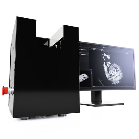
Products / High-Resolution Episcopic Microscopy (HREM)
Near-Histological Resolution in 3D for Complete Structural Insight
High-Resolution Episcopic Microscopy (HREM) 3D biomedical imaging systems for structural analysis of samples in 2D and 3D

Volumetric rendering of a cardiac sample captured using Optical HREM
What is High-Resolution Episcopic Microscopy (HREM)?
High-Resolution Episcopic Microscopy (HREM) is a 3D imaging technique for visualising samples ranging from 1-30mm. Resin mounted samples are stained, then sectioned repeatedly with the remaining block face imaged, creating high-resolution 3D stacks at 1–8-micron voxel resolution.
Advantages of High-Resolution Episcopic Microscopy (HREM)
High-Resolution Episcopic Microscopy (HREM) is a 3D imaging technique of ex-vivo samples providing multiple benefits over conventional 3D techniques such as resolution, cost and more.
Produces 1-8 micron complete voxel resolution over an entire volumetric dataset
Image dense tissue and bone at high contrast without variation
No sample clearing, distortion or interpolation
Capable of imaging up to 25mm samples with micron resolution

Whole mouse embryo captured using Optical HREM system, shown with corresponding 2D block-face section.
Flexibility for a Range of Samples
High-Resolution Episcopic Microscopy (HREM) serial section block-face imaging provides exceptional clarity and resolution, even when imaging denser tissues or tissues with varying opacities.
Unlike conventional techniques, eliminate the need for tissue clearing and interpolation, making the imaging process more efficient and convenient. HREM produces high-contrast results, enhancing the visualization of anatomical structures and details.
-
Capable of visualising samples up to 30mm in one capture or greater with scanning
-
Suitable for bone and denser tissue samples
-
High contrast images even in higher density areas
Mouse Embryos
Drosophila
Fruit & Plant Samples
Barley
Zebrafish
Other Fish Samples
Embryonic Hearts
Reproductive Systems
High Resolution for All Tissue and Sample Types
3D Biomedical imaging techniques have evolved significantly, with HREM standing out for its ability to deliver precise 3D tissue images of denser samples. High-Resolution Episcopic Microscopy (HREM) offers a variety of resolutions with scanning stages and a choice of optics. It can capture single shots or high-resolution scans at below 1 micron voxel size magnification through block-face episcopic microscopy.
With sections as thin as 1 µm, from our speciality sectioning system, HREM achieves voxel resolutions of 1-8 µm³, enabling the identification of fine details like individual nerves and blood vessels, surpassing other techniques.
-
1-8 micron voxel size resolution
-
XY scanning stage integrated into the software available for larger samples

High-resolution stitched Optical HREM Ultra image of a mouse section, revealing fine structural detail across the tissue.
Simplified Imaging Workflow with HREM
High-Resolution Episcopic Microscopy (HREM) data is readily applicable for both 2D and 3D analysis. Simple software packages like Fiji and Dragonfly can be used to generate impressive images and videos by inverting the LUT (Lookup Table).
HREM images can be manually or automatically segmented in the user-friendly 2-D image format, enabling precise measurements.
-
Data is stored as image files for easy manipulation (jpeg,tif and more)
-
No forced custom image analysis software required with subscriptions
2D Mouse Heart Sections
3D Mouse Heart Reconstruction
Applications of High-Resolution Episcopic Microscopy (HREM)
HREM has been widely used in many disciplines but can be extended to a range of morphological studies. Imaging denser samples up to 25mm down to below 1mm with voxel sizes down to 1 micron. It is most commonly used in samples from 1mm-12mm cubed.
Mouse Embryos
HREM has been widely used to image full mouse embryos across all stages of development
-
Full 3D reconstruction of whole mouse embryos in high detail
-
Track developmental abnormalities across gestational stages
-
Quantify organ formation and spatial relationships
-
Ideal for genetic and transgenic studies
Plant Samples/Seeds
-
3D imaging of seed structures and root development
-
Image vascular bundles, meristems and embryogenesis
-
Ideal for agricultural trait research and seed phenotyping
Mouse Hearts
HREM has aided mouse heart imaging at all stages.
-
Visualise intricate 3D cardiac structures and vasculature
-
Study congenital heart defects (CHD) and structural remodelling
-
Ideal for phenotyping mutant mouse lines
Small Animal Organs
-
Capture kidney, liver and lung in full 3D under 1 cm
-
Measure volume, surface area and branching networks
Chick Embryos
-
Large scale imaging of early vertebrate development
-
Assess segmentation, heart looping and organogenesis
-
Bridge developmental biology with anatomical analysis
Optical HREM Systems
We offer two main Optical HREM system solutions, available worldwide, offering flexibility in budget and in capture. Each systems can achieve highly detailed 3D image stacks, while allowing the systems to be accessible to more.
.jpg)
OHREM Ultra
Ultra OHREM systems offer flexible imaging with XY scanning stages for up to 60mm with various illumination and optical setups.
-
~60 x 60 x 30 field of view, with scanning
-
Choice of upright or zoom optics
-
Ideal for fluorescence applications

OHREM Micro
Micro OHREM Systems offer simple imaging of samples up to 25mm in FOV, featuring single shot optics.
-
25 x 20 x 20 field of view
-
Simplistic single shot imaging
-
Smaller form factor
Contact our High-Resolution Episcopic Microscopy (HREM) Experts
Want to know more about HREM, ask for a quote or get questions answered. Contact us and we can help answer all your questions.
Phone:
+44(0) 1462633500
Email:
hello@indigo-scientific.co.uk