High-Resolution Heart Imaging in Mice and Embryos with Episcopic Microscopy (HREM)
High-Resolution Episcopic Microscopy (HREM) provides detailed 3D imaging of heart structures in mice, mice embryos, and chicken embryos, advancing cardiovascular research
Visualise precise 3D visualization of cardiac structures in mice, mice embryos, and chicken embryos with HREM with samples ranging under 1mm up to 25mm.
3D Heart Imaging in Small Models with HREM
High-Resolution Episcopic Microscopy (HREM) is a biomedical imaging technique that gives insights into the cardiac structures of small models such as mice and embryos. Whether you're studying the developing hearts of mice embryos or chicken embryos, HREM delivers the clarity and detail required to advance your cardiovascular research. By producing high-resolution, 3D images, this technology allows researchers to explore the complex anatomy of the heart in different species.

Applications of HREM in Cardiac Research
HREM allows researchers study heart development and disease in animal models. Here are key applications:
-
Heart Development in Mice: HREM enables detailed 3D imaging of heart development in mice, offering critical insights into congenital heart.
-
Mice Embryo Cardiac Imaging: This technique allows for precise imaging of cardiac structures in mice embryos by imaging the full embryo, making it possible to study the heart at various stages of embryonic development.
-
Chicken Embryo Heart Analysis: HREM is also effective for imaging chicken embryos, providing valuable data for comparative studies in cardiac research.
-
Larger Samples: HREM can be used to image denser larger cardiac systems, such as pups
How High-Resolution Episcopic Microscopy Enhances Heart Imaging
HREM offers a unique approach to imaging heart structures by producing high-resolution, 3D volumetric data. The process involves embedding the specimen, sectioning it, and capturing episcopic images, which are then reconstructed into a 3D model. This method is particularly beneficial for:
-
High-Throughput Imaging: HREM allows for detailed imaging in quantity allowing for knockout trials.
-
Detailed Visualization: The technique provides a clear view of the intricate structures of the heart, including valves, chambers, and vessels for further study.
-
Quantitative Analysis: HREM enables precise measurement in 3D ow High-Resolution Episcopic Microscopy Enhances Heart Imaging and analysis of heart structures, which is critical for understanding heart development and disease.
-
Do you ship globally?Yes we ship and service OHREM's globally, service contracts are provided to help maintain your machine.
-
Can a demo unit be organised at my institution?Indigo is a small company and uses its size to give the best possible product for the best possible price to help researches get the most from their product. To keep costs low we do not have a facility to deliver demo stock to customers. Instead we invite customers to make specimens and send them to us to either be processed by us or one of our existing customer network. Get in contact to see how we can help.
-
What is the difference between HREM and OHREMHREM and OHREM are interchangeable however at Indigo we refer to OHREM as the system and HREM as the Technique. OHREM stands for Optical High Resolution Episcopic Microscopy.
-
Can the machine be adapted to different techniques?The system is flexible and we can help adapt the system to user requirements, get in contact to see what we can do.
-
Who Manufactures OHREM?The Optical HREM System is manufactured and sold by us here at Indigo Scientific.
-
How is the OHREM controlled?OHREM is controlled by a custom application designed by us here at Indigo to help deliver the most optimal performance and efficiency.
-
What is the maximum size of a specimen?A single shot optic (no scanning) is limited to the optical limit of 30mm for the standard system. However, with changes to the configuration the OHREM can achieve success of samples of 60mm wide. Get in touch to find out more.
-
What samples can be used?In theory it is down to the staining but already there has been great success in a variety of different specimens, we can offer interested parties imaging of there samples after following the protocol found in the members area. Get in touch to find out more.
-
How does the system (OHREM) work?HREM works by using a custom made microtone and optical setup made by Indigo Scientific to section a sample. The sample is imaged during sectioning giving an image series. Learn more about High Resolution Episcopic Microscopy Here. HREM sections 1-10 microns of a resin based block and images the surface.
-
How many samples/blocks can be imaged at once?Revision 1 OHREM systems can only image 1 block per section. OHREM 2 and an adapted OHREM 1 is capable of imaging 2 20 mm blocks or 4 10 mm blocks with the facility to expand this if required. It is worth noting a block can contain more than one sample, for example one block can contain 3 embryonic mouse hearts.
-
Are there different configurations/Addons?OHREM comes with many addons and configurations. Selections can include a scanning stage, dual/multi flourescence etc, contact us to get full details.
-
How does the system look?The Optical HREM system will vary based on the type of system you want but the main system will consist of a microtome unit and optics. The two major systems are the HREM Ultra and HREM Micro .
-
Can HREM be used for imaging hearts at different developmental stages?Yes, HREM is highly effective for imaging hearts at various stages of development, particularly in embryos and pups.
-
What makes HREM a good tool for heart imaging in small models?HREM provides high resolution and 3D imaging capabilities, allowing researchers to visualize the complex anatomy of the heart in great detail and measure in 3 dimensions.
-
Can HREM be used to image other species and organs?Yes, HREM can be used to image many different sample types and density’s such as zebrafish.
-
What are key features to look for in a microscope camera?There are lots of specifications, options and software that would impact your choice of a microscope camera. To keep things simple let’s point out some key features we would advise you look out for Sensor size – In microscopy you want to look out for the sensor size, a larger sensor size (often the biggest seen at 1-1.1 inches) often allows for greater pixels which gives better sensitivity for low light applications. It may provide greater resolution up to 20 megapixels. For most industrial and lab work applications a trade off of sensor size over cost is usually taken, picking a smaller sensor size with a fair pixel size. Pixel size – Pixel size refers to the size of each individual pixel on the sensor, the greater the pixel size the better it is for low light applications. The lower the pixel width and height the generally more noisier the image, with lower dynamic range but can often fit far more pixels on the same sensor. Colour type – Microscope cameras come in two main colour variants (not depth that’s a different thing), colour and monochrome. Monochrome cameras are best for low light applications where you require better depth and sensitivity over being able to see the sample in colour. Most applications(bright field etc) will want a colour camera. Don’t be shocked that monochrome cameras carry a bigger price tag either, they are often designed for fluorescence imaging so have better specifications. Interface – Believe it or not picking a camera with a good interface is important. For example USB 3.0 is a well known supported interface, Ethernet is also good but may be more complicated for plug and play scenarios. Some cameras require external power which increases cables. Ultimately consider a camera that meets these requirements, we would highly recommend a USB camera with no external source required. Software for Image Analysis and Capture – Choosing a camera can also be a choice of software package, make sure you weigh up licenses, functions, future upgradability and compatibility with your favourite packages such as micromanager. That’s a good start for you to get choosing, if you need any specific help contact us.
-
How Do Colour Microscope Cameras Work vs Monochrome Microscope Cameras?Traditional colour microscope cameras use a red, green and blue filter (in varying formats) and filters incoming light to given pixels. Each pixel will have one colour component and then interpolation is used to give the final full colour image. Monochrome cameras simply measures the intensity of light, this is across all of the spectrum.
-
What are the main specification differences between Colour Microscope Cameras and Monochrome Microscope Cameras?

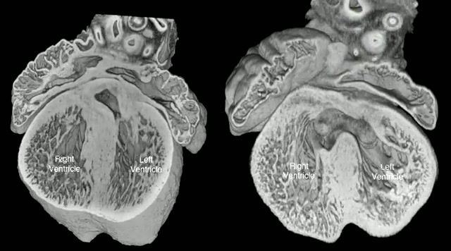
Gallery
Below is a gallery of HREM example images captured by ourselves and our customer base of Cardiovascular examples.


Add HREM to your Research Tools for Cardiac Research
Contact us today to learn more about how High-Resolution Episcopic Microscopy can enhance your studies of heart development in mice and embryos.
Phone:
+44(0) 1462633500
Email:
hello@indigo-scientific.co.uk